Enhancing usability and improving examination efficiency with optical camera. Release of RADspeed Pro SR5 general radiographic system.
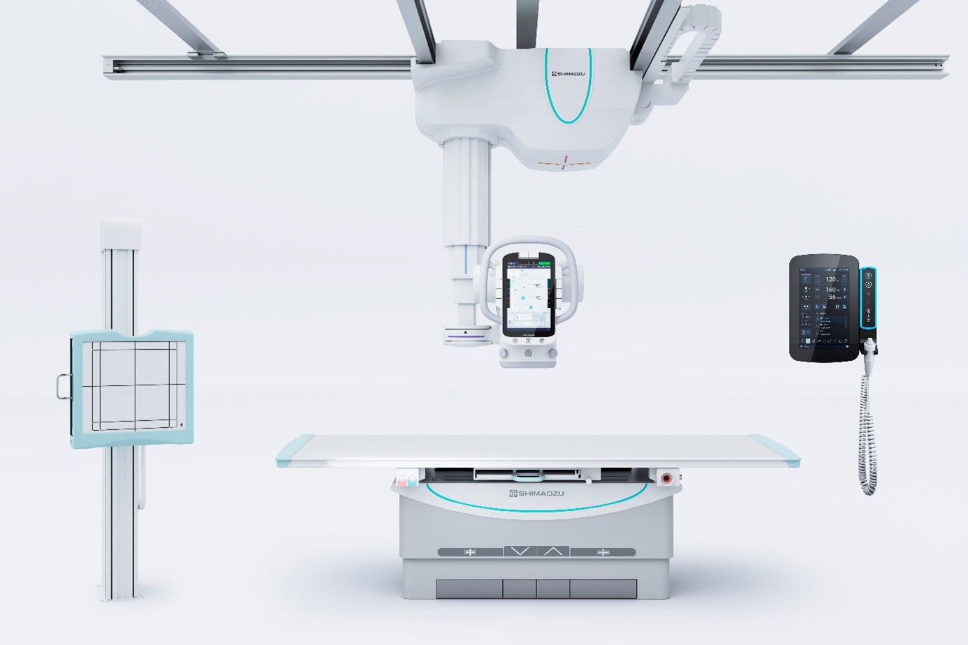
RADspeed Pro SR5 digital general radiographic system
Introduction of new RADspeed Pro SR5
Presentation of new RADspeed Pro SR5
Shimadzu Corporation has released the general X-ray system RADspeed Pro SR5. This product aims to improve usability through enhancements in the console and the inclusion of an optical camera that detects patient movements. Those features improve the efficiency of examination procedures and reduce the burden on both healthcare workers and patients.
The RADspeed Pro SR5 Version is a General Radiographic System used for imaging various parts of the body, including the head, chest, abdomen, and limbs. As the workload increases due to infection control measures, there is a demand for features that allow healthcare workers to efficiently perform examinations while focusing on the patient.
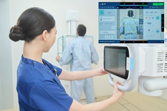
The new model includes completely redesigned controls for performing radiography using a console with easier operability and better visibility. It has also improved the handle shape of the X-ray tube support for easier gripping. The “VISION SUPPORT” imaging assistance function (optional), which mounts an optical camera to the X-ray irradiation part, allows the overlay of the patient’s image on the monitor with the X-ray irradiation area and the position of the X-ray detector. This supports the smooth determination of imaging positions, which is usually a time-consuming task. If the patient moves from the determined position, the optical camera detects the movement and notifies with sound and display, reducing the risk of retakes.
Example of new camera supported X-ray examination
Shimadzu’s medium-term management plan specifies implementing a strategy referred to as “imaging transformation” (IMX) for combining AI or IoT technologies with diagnostic imaging equipment to provide new added value. Shimadzu will continue to focus on automation in General Radiographic System through AI technology, aiming to contribute to “human life and well-being”.
Features
1. Enhancing examination efficiency and reducing the burden on healthcare workers
The control console of the X-ray tube support has been equipped with a large touch panel monitor, and the display of the X-ray generator's operation console has been improved, enhancing operability and visibility. The handle of the X-ray tube support has been designed to be easy to grip and operate. The wireless remote control* for automatic tube positioning offers additional operating convenience. The new wireless hand switch* with color-coded status display offers maximum flexibility and safety during exposure. (* optional)
2. Positioning Assistance with Camera
“VISION SUPPORT” imaging assistance function was developed for assisting examinations based on camera images.
By displaying the camera image of the patient, the X-ray exposure area and additional information on the monitor of the tube stand and the X-ray generator, the operator can visually check on the monitor whether the patient is within the exposure area.
VISION SUPPORT can also display the position of the X-ray detector and the automatic exposure field chambers to enable more accurate positioning. As these images can be reviewed both in the examination room and in the control room, examinations can be carried out while concentrating even more on the patient than before.
On the monitor of the X-ray stand and the control panel of the X-ray generator, it is possible to overlay the patient image and the X-ray exposure area allowing a visual confirmation of whether the target area is within the exposure area.
3. Rich Applications Using Camera
For VISION SUPPORT, an extensive selection of application software is available for using the camera images to assist users. With "Motion Detection", the system displays the subject’s body movement on the screen from the predetermined imaging position and it provides both visual alerts and sound warnings, if movements exceed a certain threshold. There is also an optional "Last Position Display" which allows switching between the previously captured image and the real-time image. Furthermore, it is possible to transfer camera image to a digital radiographic system for image processing. During patient positioning, the DR system displays and analyzes the video image to compare it with the selected protocol, which can reduce positioning errors*.
• Note: Only in combination with CXDI-DR system. VISION SUPPORT is a trademark of Shimadzu Corporation.
For more details, visit RADspeed Pro SR5 .