RADspeed Pro™ SR5 Version
Features
Optical camera application creates an environment where medical personnel can focus on patients
Vision for patient concentration
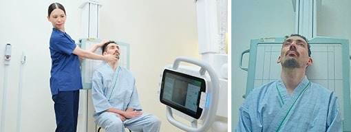
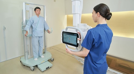
The video image from a camera built into the collimator is displayed on the X-ray tube support control panel and high-voltage generator control panel monitors. The optical camera application provides an environment where medical personnel can focus on patient care.
Supporting you watching over patients
Equipped with optical camera
In addition to visually checking on patients, you can observe patients from both the examination room and control room.


Reduces positioning effort and improves accuracy
Live view display
Supports accurate positioning by showing overlay of detector area, irradiation field and AEC pickup fields*, which are difficult to check directly.

*Guide line overlay is for reference only.
*AEC pickup fields overlay is not available in the United States.
Reduces frequency of repeating exposures due to body movement
Motion detection
Patient body movement can be confirmed from the point that body movement detection mode is activated.*

*Check the patient's condition, even directly visually.
Smoother positioning correction during repeated exposures
Last position display
By checking the immediately previous exposure positioning, positioning can be achieved more smoothly when repeating exposures.
Innovations that improve operability
Vision for easy operation
Healthcare requires multiple complex tasks. To support this hectic work, it is essential to achieve an examination environment that contributes to diagnosing patients, while also ensuring simple and intuitive operability. Shimadzu offers systems optimized for usability.
Displays equipment status
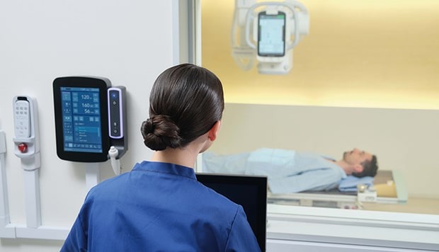
Illumination improves visibility 
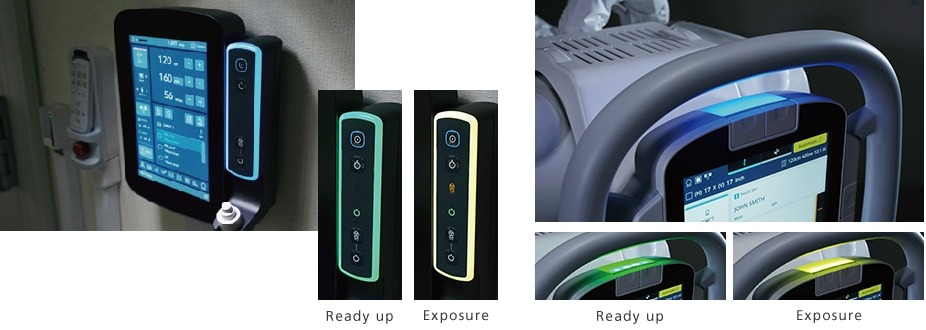
Illumination of the X-ray high-voltage generator and ceiling-mounted X-ray tube support enables better understanding of the instrument status. In addition, the hand switch illuminates to indicate the system is ready for the next exposure.
Easy to operate in high positions
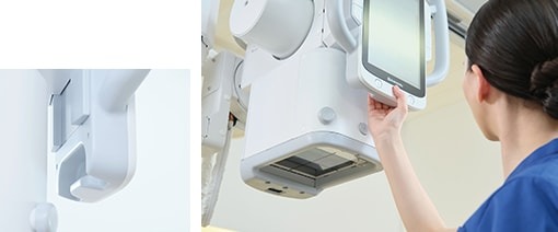
Lower hand grip 
A hand grip is provided on the back side at the bottom of the control panel, and operation is possible by pressing the all-free switch on the front side. Operation is easy even when the X-ray tube support is located in a high position.
Intuitive operation
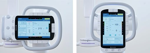
Graphic display of unlock buttons 
Graphic unlock buttons enable more intuitive operability by displaying button symbols with the unlock direction oriented to match the perspective of the operator in either supine or standing positions.
Assist functions support positioning
Vision for reduction of operator burden
A wide range of positioning support functions reduces the operator burden.
Superb operability
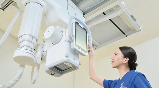
Power assist function OPTION
Motors assist handle operations. This reduces the burden on operators during movements by enabling the ceiling-mounted X-ray tube support to be moved quickly and lightly.
Changeable with one touch
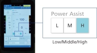
Assist level adjustment
Large movements can be made quickly with lighter force, and precision movements can be made for detailed positioning.
Positioning assistance with remote control
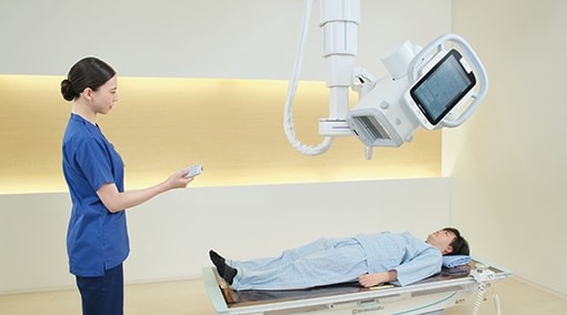
Auto-positioning feature OPTION
The X-ray tube support can be moved by remote control. This enables smooth positioning while observing the patient.
No cables in the way
Wireless remote controller for automatic positioning
An infrared wireless remote controller is used to prevent cable interference. In addition to instrument movements, it can also control the collimator. Actions immediately stop when the remote control operations are stopped.

High throughput
Vision for high throughput
Use it to perform examinations smoothly, while relieving patient anxiety. To achieve both, a system is required that can shorten examination times while ensuring safety. Shimadzu supports efficient examination process flows for front-line healthcare workplaces.

Auto stitching of long view images
Speed stitch function OPTION 
The X-ray tube swings and the FPD moves automatically to capture images.The captured image data is then automatically stitched together in the DR system. That makes it easy to create images that are wide along the longitudinal direction of the body.*
- *This functionality is available for systems that combine a Shimadzu BR-120 or BR-120T Bucky stand and a BK-200 Bucky table with a DR system from other manufacturers. For information about compatible DR systems, contact a Shimadzu sales representative.
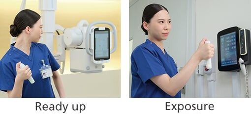
Operable from any position on the operation room
Wireless exposure switch OPTION 
A Bluetooth wireless hand switch enables freely acquiring images from any position in the control room.*
- *There are restrictions on available countries.
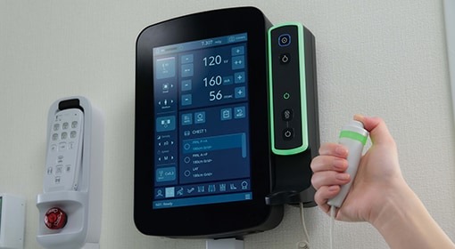
600 kHU quick ready X-ray tube
Speed shot function OPTION 
Because only 0.8 seconds is required to prepare for exposures after pressing the exposure button, images can be acquired quickly, even for patients with difficulty holding their breath or holding a particular body position. The X-ray tube anode starts high-speed rotation when the collimator lamp is illuminated.
Consideration for the patients
Vision for patient care
Patient care in physical form.
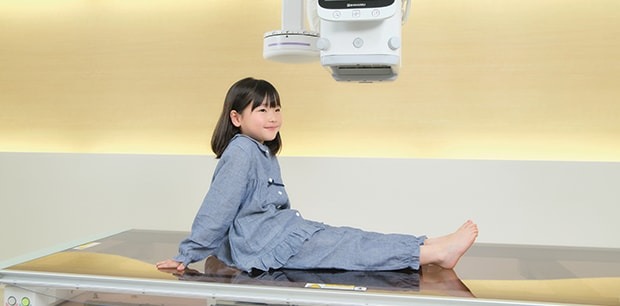
Rubber Cushioning for
Extra safety 
The bottom of the X-ray tube support and the perimeter of the collimator radiation port are covered with soft rubber cushioning material. That tenderly protects patients by reducing their risk of injury from unexpectedly sitting up after exposures have been taken in the supine position and hitting their head on the instrument.
Reduction of radiation exposure
Collimator with automatic filter 
An automatic filter function is included that automatically switches the filter coupled with the collimator when the APR mode is selected based on the exposure area. Four filter modes can be preset (0.1, 0.2, or 0.3 mm thick copper or no filter).
Smooth patient confirmation
Check patient information in the examination room OPTION 
Patient information can be displayed on the control panel. This ensures patients can be smoothly identified in the examination room.
Department of Radiology, Fukuoka University Hospital
Department of Radiology, Japanese Red Cross Aichi Medical Center Nagoya Daini Hospital
(formerly Japanese Red Cross Nagoya Daini Hospital)
Global Marketing Department, Medical Systems Division, Shimadzu Corporation1
Research & Development Department, Medical Systems Division, Shimadzu Corporation2
Medical Systems Division, Shimadzu Corporation
Department of Radiology, Diagnostic Imaging Center
Osaka General Hospital of West Japan Railway Company
Division of Radiology, The University of Tokyo Hospital


