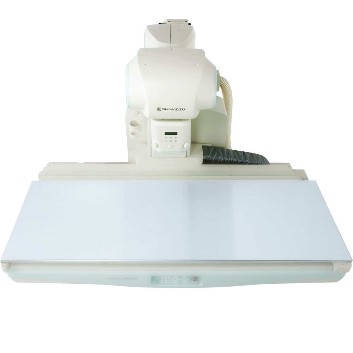
Sonialvision G4 LX edition
Digital Multipurpose R/F System
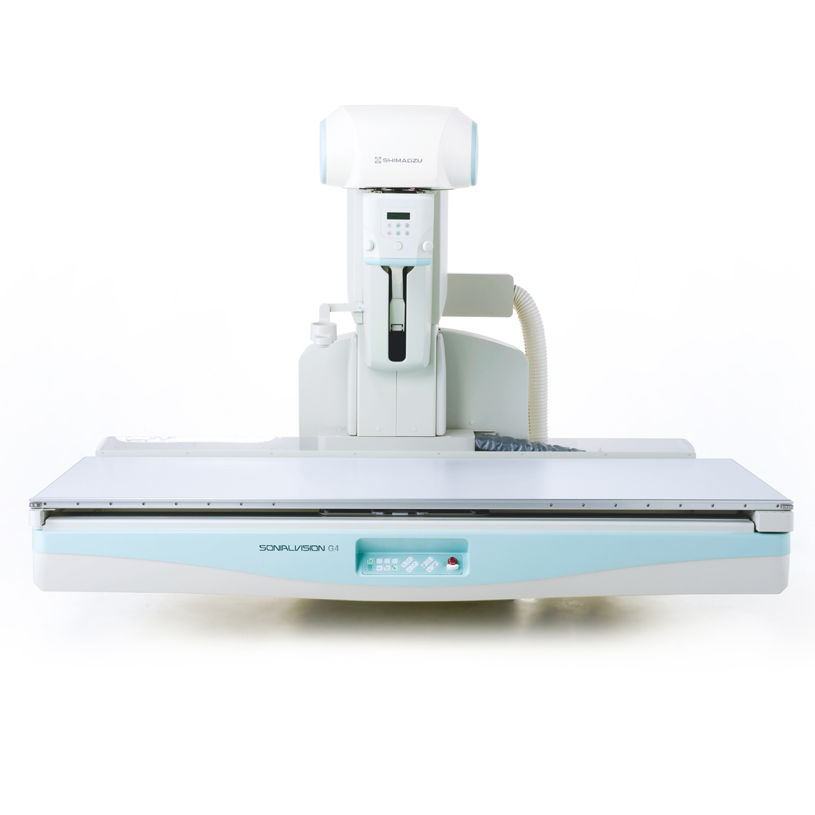
The Sonialvision G4 LX edition is a premium R/F system with outstanding functionality and quality for a wide range of examinations. As a universal system, it meets the individual and high requirements of imaging departments and institutes.
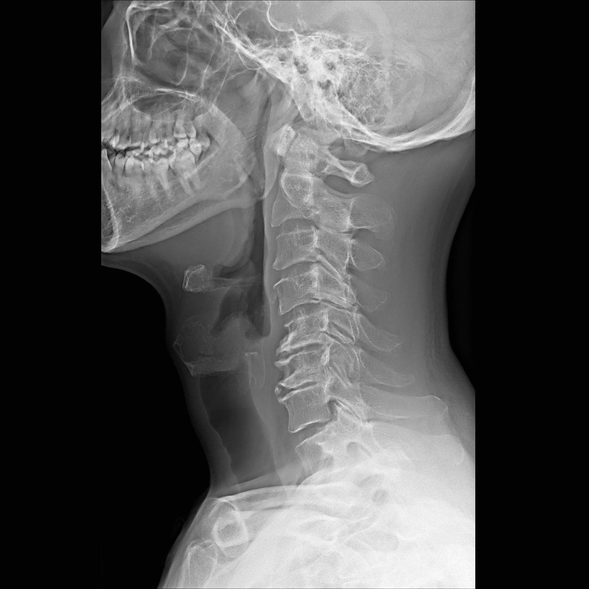
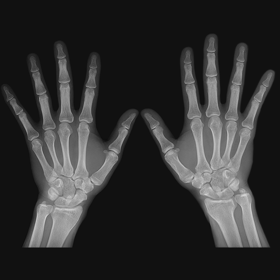
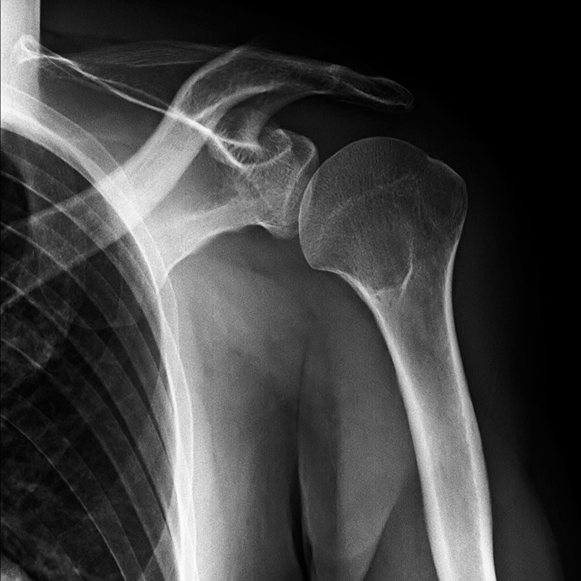
Ultra-high definition and dynamic images
The dynamic detector has a small, 139 μm pixel pitch and a high-sensitive CsI scintillator. Combined with the SUREengine-Advance and SCORE PRO Advance it ensures clearer fluoroscopy and radiography images. The FPD offers five sizes of selectable fields of view up to 43 x 43 cm, allowing a wide range of examinations.
Comprehensive dose reduction and management

Equipped with various functions to reduce radiation exposure levels effectively, the system provides peace of mind for both patients and attending personnel during examinations. Highly developed mechanisms for efficient dose reduction and X-ray exposure control are available.
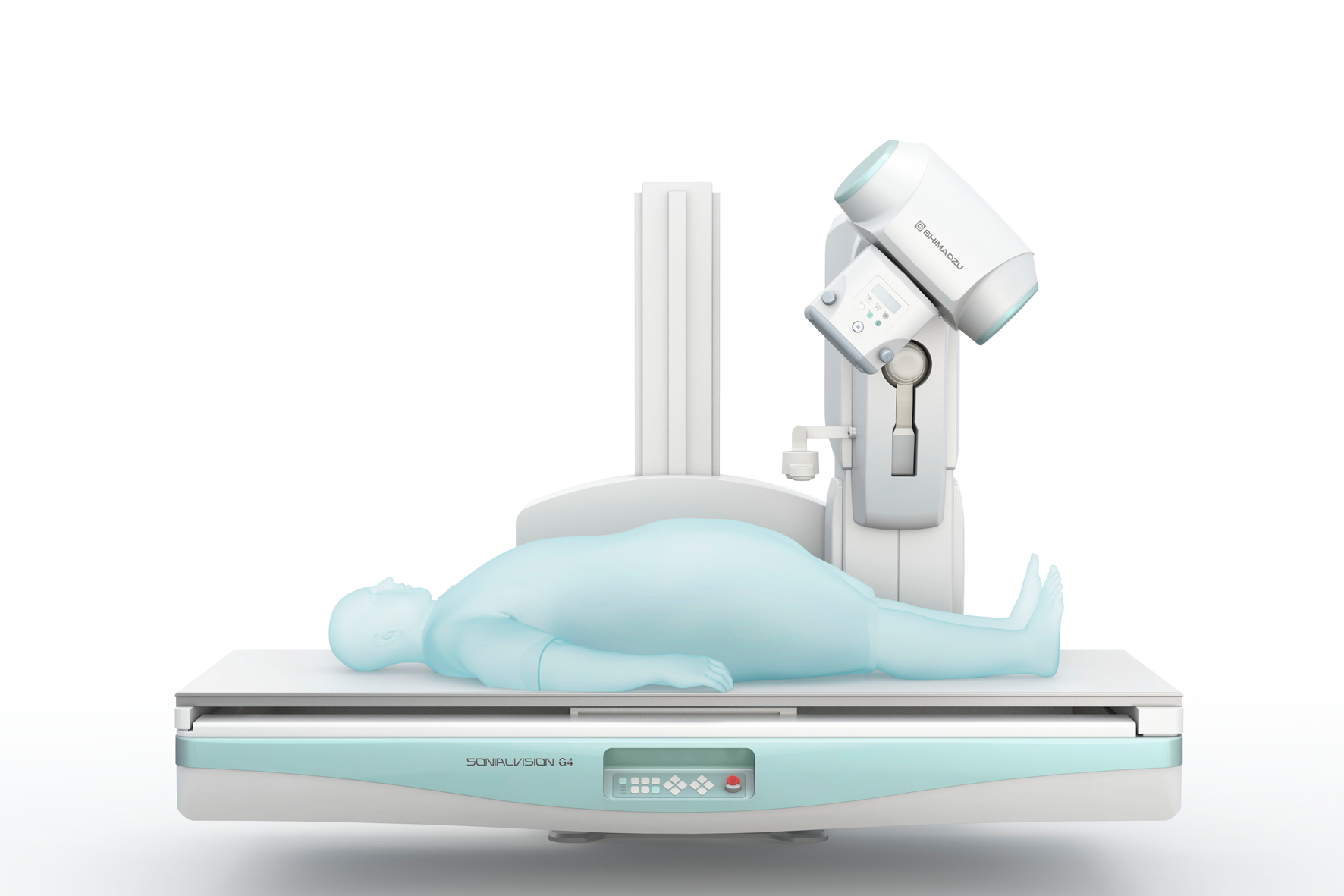
Sonialvision G4 LX edition X-ray table offers advanced functionality
Compact system
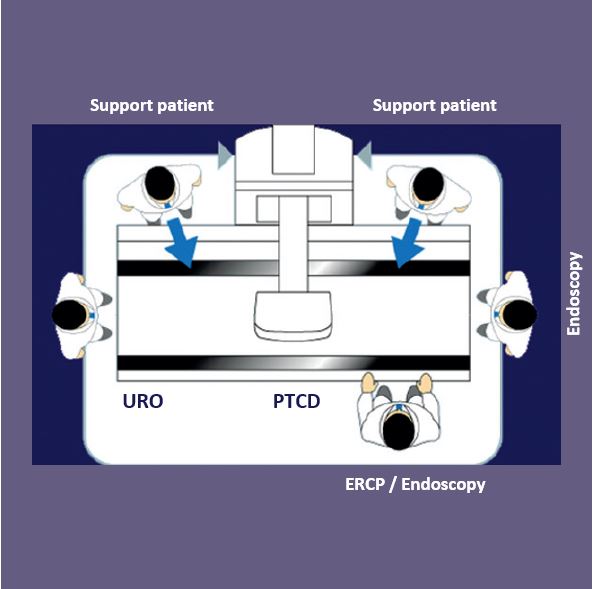
It allows access from all sides improving patient support for a wide range of examinations.
Wide coverage and fast positioning
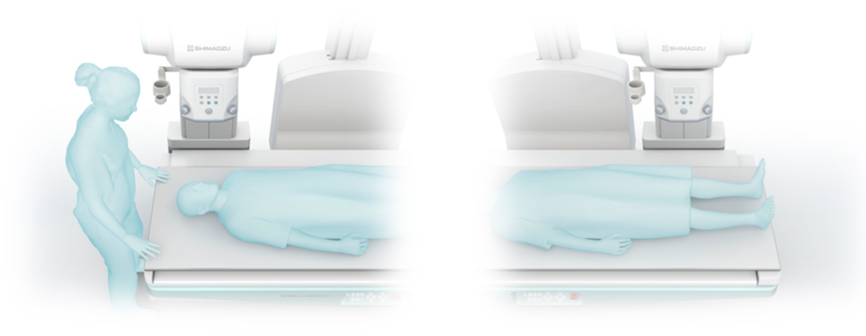
The wide longitudinal movement range captures the entire body without moving the patient.
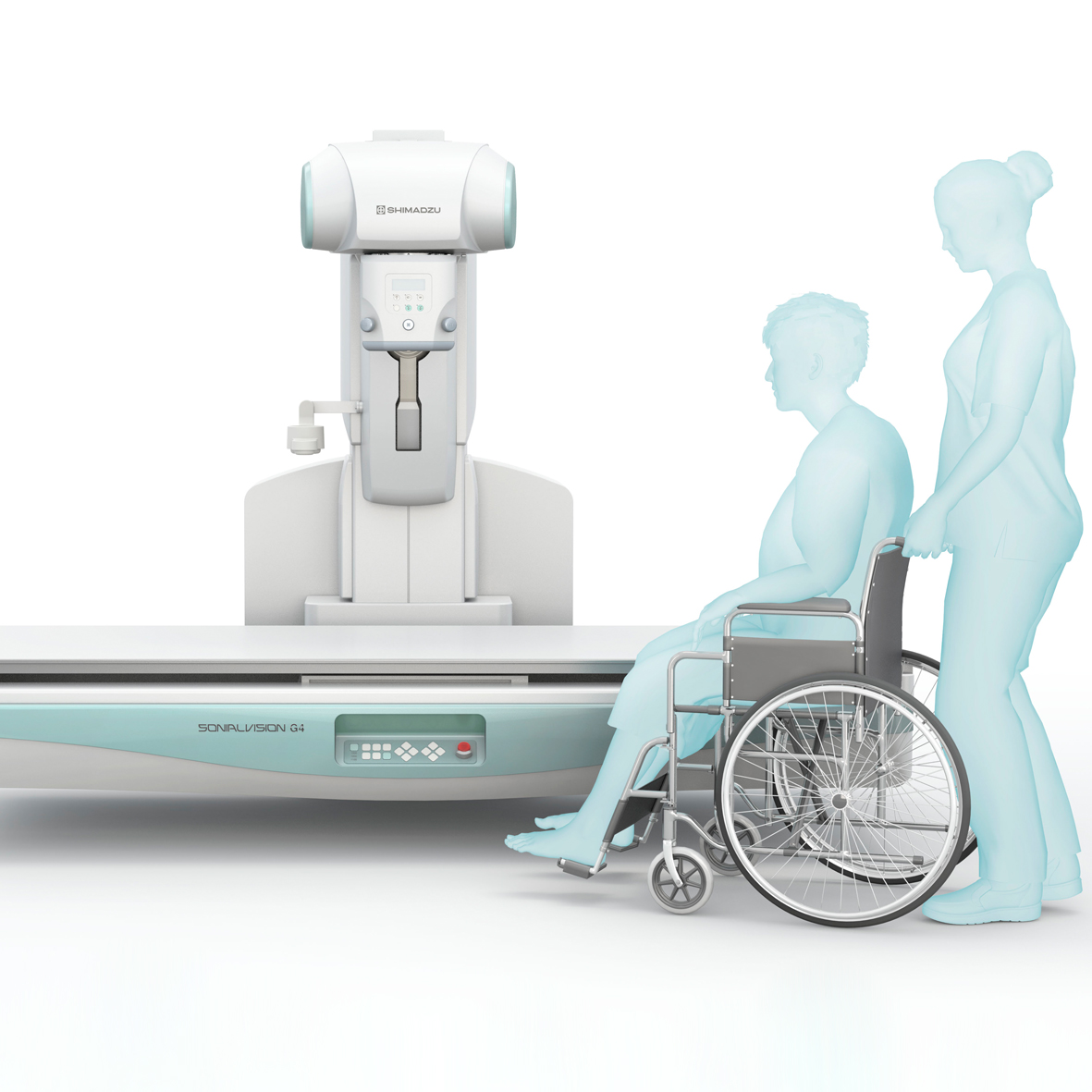
High patient load capacity
The wide, clean, seamless table offers the highest weight capacity easily accommodating bariatric patients as well.
Table height adjustment
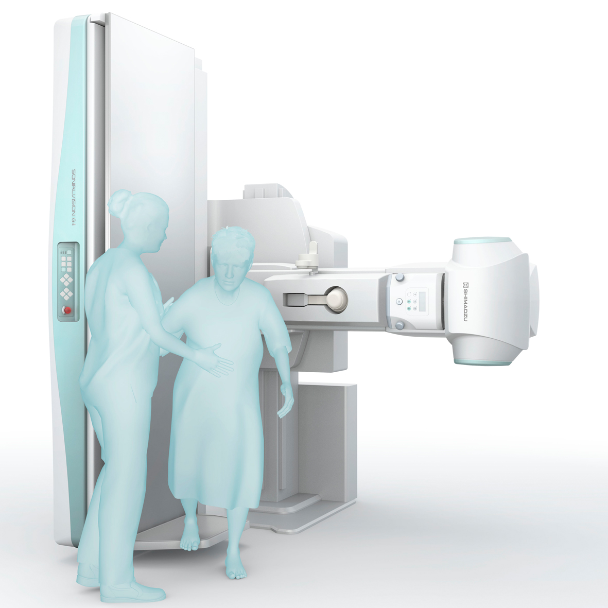
Flexible height positioning of the seamless tabletop provides ideal hygienic and working conditions as well as easy and safe access for any patient.
Advanced imaging technologies increase efficiency

The Sonialvision G4 2-in-1 R/F system performs
- digital radiographic
- pulsed-fluoroscopy
- extended video-fluoroscopy examinations
and additional advanced imaging technologies, like
- DSA
- real-time and motion-tolerant RSM-DSA
- Slot Radiography and
- Tomosynthesis for general radiography examinations.
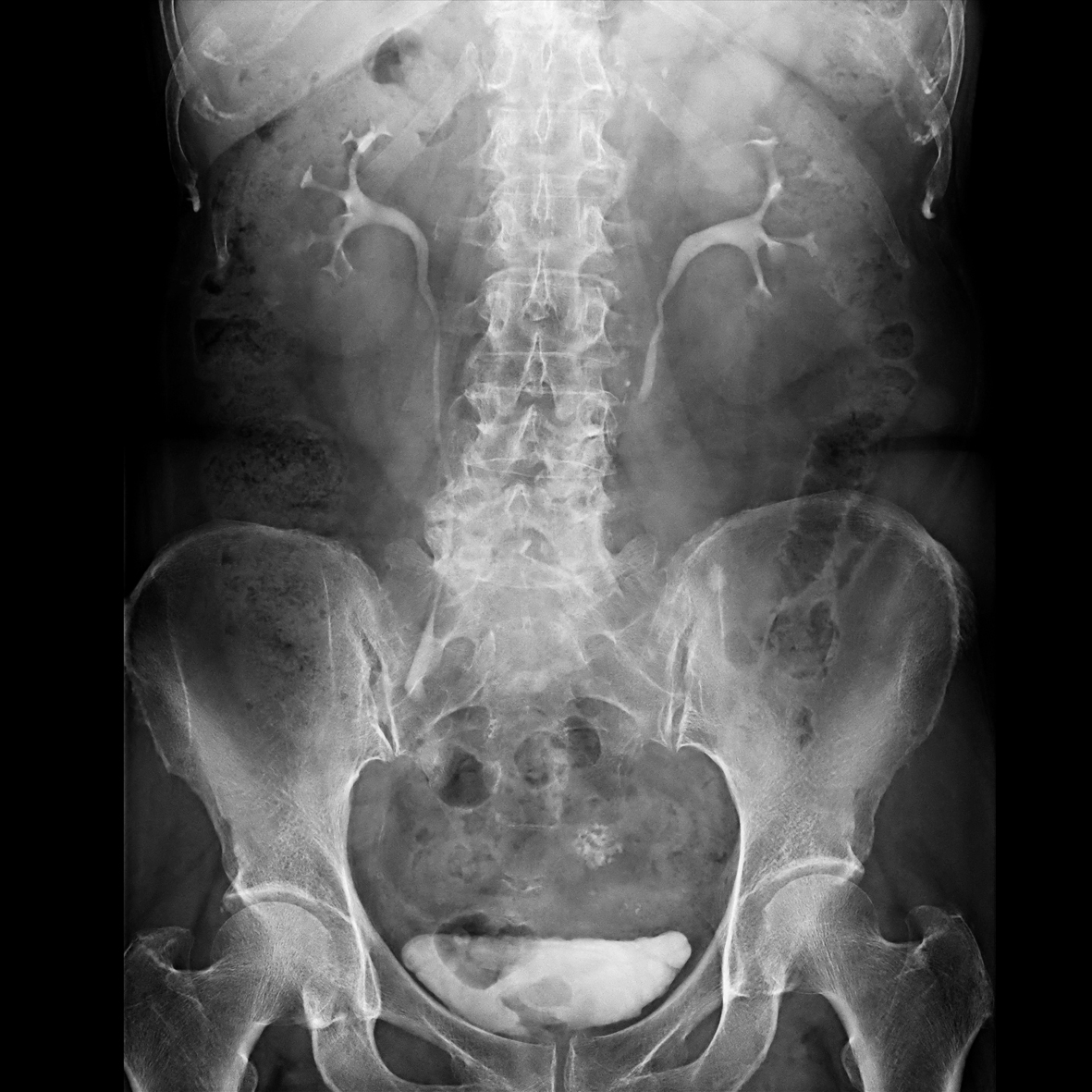
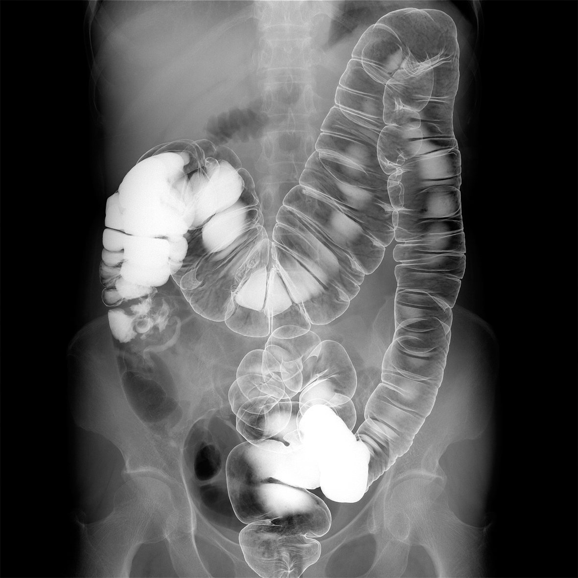
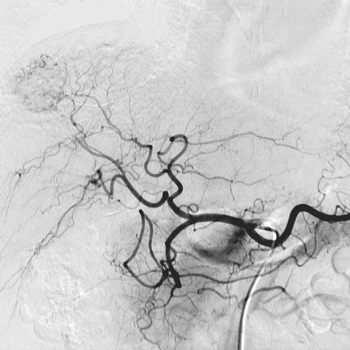
DSA & real-time and motion-tolerant RSM-DSA
The large field of view means that DSA can be used for examinations of the hepatic artery to the entire lower extremities. The RSM-DSA filter provides continuous automatic masking resulting in a DSA-like image even when the patient or the table is moved. It produces excellent results where patient movement is inevitable, such as bolus chasing and abdominal examinations, particularly when patients find it difficult to hold their breath.
Slot Radiography
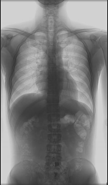
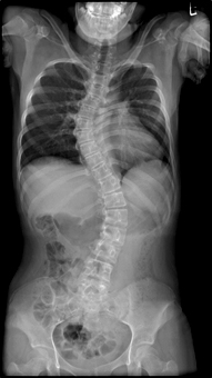
Slot Radiography provides a quick and efficient way to acquire long leg and full spine images. Images can be acquired with the table in any position (from supine to erect) making weight-bearing procedures easy. The large FPD and the slit beam exposure method results in better patient coverage than traditionally achievable with CR cassettes. In addition, the slot technique produces low dose images with minimal geometric distortion and accurate measurements.
Tomosynthesis
This function is an automated acquisition process that acquires several images while the tube is moving in an arc across the region of interest, similar to the more conventional topographic movement. The series of images is then processed in a “cone beam” or CT-type back projector resulting in a volume of data available for investigation by scrolling through it in a series of slices. Slice thickness can be post-processed in a similar manner to that used in CT imaging. Slices of interest can be extracted as required for further investigation, printing and DICOM storage.
Tomosynthesis provides numerous key benefits including:
- low-dose
- weight bearing
- low metal artefacts
- high spatial resolution
- high quality imaging in plaster fixation
- easy positioning
- fast acquisition
- large field of view (FOV)
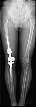
Standing position tomosynthesis image of weight-loaded knee joints
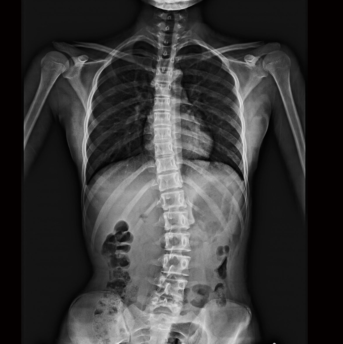
Precise fracture diagnosis (Courtesy of Okitama Public General Hospital)
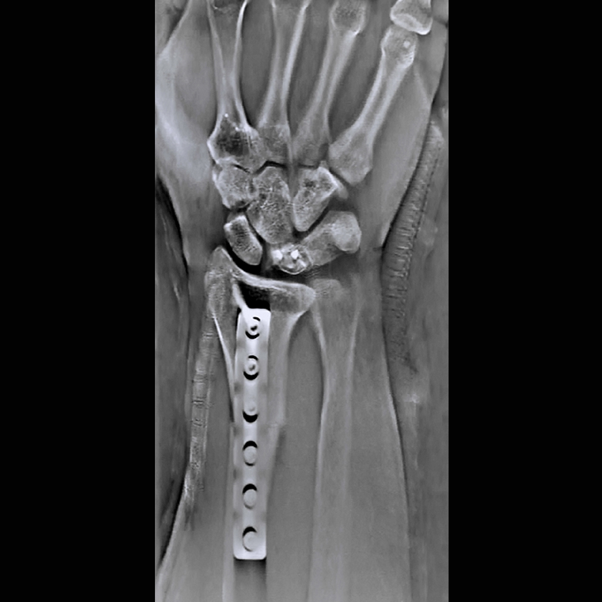
Clear visualization for callus formation (Courtesy of Nara City Hospital)