SLOT Advance
Ideal technology for long-view images with maximum accuracy
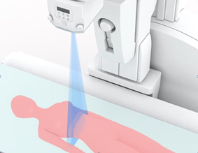
With just a simple workflow, SLOT Advance provides high accuracy images with long fields of view, such as for full spine or full leg images, taken with minimal X-ray dose. SLOT Advance acquires a series of accurate images of a few centimetres central slit as the imaging chain moves successively along the patient.
Providing super high accuracy measurements SLOT Advance collimates the X-ray beam, exposing just a narrow central slit field without the distortion caused by oblique rays. These central slit images are captured using the best-in-class SONIALVISION R/F table’s super-high resolution Flat Panel Detector (FPD) by moving it in parallel with the X-ray tube. These successive images are reconstructed automatically to create one long image in SONIALVISION G4’s digital imaging unit (DR-300). This simple workflow enables highly accurate measurements without distortion.
Significant reduction of examination time
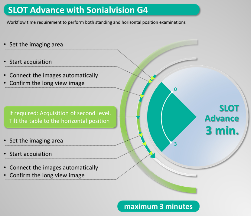
Before starting the exposure, the start and end positions of the examination field are simply set. That’s all it takes to obtain proper long-view images using Slot Radiography. All post-processing required to connect, adjust, and display the images on the monitor is done automatically immediately after exposure. SLOT Advance eliminates the time-consuming steps of setting up the cassette and making adjustments, not to mention moving the patient between standing and horizontal positions, all of which are required by conventional CR long-length imaging.
High image quality with minimal dose
Due to the SONIALVISION G4’s super-high resolution FPD, SLOT Advance simultaneously achieves both higher image quality and lower dose levels. The flat panel detector features the smallest pixel size in its class of 139 µm, and Shimadzu’s advanced imaging technology improves contrast resolution.
Extra-wide imaging range
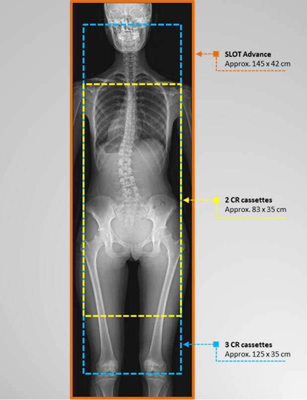
The combination of SONIALVISION G4’s extra-long imaging chain slide coverage, and its large FPD of 43 cm (17”) achieves even a wider longitudinal and transverse imaging area (up to 145 x 42 cm) compared to the image stitching capabilities of CR units.